🧪 Cerebrospinal Fluid
Production, circulation, composition, and the essential clinical tests — a complete study guide.
CSF Production & Circulation
Cerebrospinal fluid (CSF) is a clear, colourless fluid that fills the ventricular system of the brain and the subarachnoid space surrounding the brain and spinal cord. It serves three vital functions: mechanical cushioning (a buoyant shock absorber for the brain), chemical homeostasis (maintaining the ideal ionic environment for neuronal function), and waste clearance (removing metabolic by-products).
The Choroid Plexus — The Main Factory
Approximately 70% of CSF is actively secreted by the ependymal cells of the choroid plexus. These highly vascular tufts of specialised epithelium are found hanging from the roof of the lateral, third, and fourth ventricles. They act like a specialised kidney, selectively filtering plasma from the blood and secreting it as CSF. The remaining 30% comes from cerebral blood vessels and the ependymal lining of the ventricles themselves.
CSF Production Stats
The numbers are worth memorising — they come up in clinical scenarios and MCQs:
Total volume: ~150 mL (130–150 mL) in the entire CNS; only 30–40 mL is actually within the ventricles at any one time.
Production rate: ~500 mL/day (0.3 mL/min).
This means the entire CSF volume is completely replaced approximately 3–4 times per day. Any impairment of drainage therefore causes rapid pressure build-up.
The ventricular system is a series of interconnected, CSF-filled cavities deep within the brain. CSF flows in a strict, one-way sequence — any obstruction at any point causes the system upstream to balloon with trapped fluid.
Lateral Ventricles (×2)
The journey starts here. Each cerebral hemisphere contains a C-shaped lateral ventricle with anterior (frontal), posterior (occipital), and inferior (temporal) horns. The choroid plexus here is the largest and most productive. This is where the bulk of CSF is made.
Foramina of Monro (Interventricular Foramina)
CSF flows medially from each lateral ventricle through these paired openings — one on each side — into the single midline third ventricle. A tumour near the Foramen of Monro (e.g. a colloid cyst) can cause obstructive hydrocephalus with sudden postural headache — classic exam scenario.
Third Ventricle
A narrow, slit-like midline cavity nestled between the two thalami (in the diencephalon). The walls are formed by the thalamus on each side. The pituitary stalk passes through its floor. The choroid plexus also runs in its roof.
Aqueduct of Sylvius (Cerebral Aqueduct)
The narrowest point in the entire CSF pathway. This tiny channel (~1.5 mm wide) tunnels through the midbrain (mesencephalon) to connect the 3rd and 4th ventricles. Aqueductal stenosis is the most common cause of non-communicating hydrocephalus — dilation of the lateral and 3rd ventricles with a normal 4th ventricle is the CT hallmark.
Fourth Ventricle
A diamond-shaped cavity sitting anterior to the cerebellum (the cerebellar vermis forms its roof), and posterior to the pons and upper medulla. The floor is the “rhomboid fossa” — an important surgical landmark containing cranial nerve nuclei. This is the last stop before CSF exits the internal system.
Exit Apertures — Leaving the Ventricles
CSF exits the 4th ventricle through three openings in its roof (see the M&L mnemonic below). It then enters the subarachnoid space to bathe the exterior of the brain and spinal cord.
Subarachnoid Space & Cisterns
Once outside the ventricles, CSF flows through the basal cisterns (large subarachnoid reservoirs at the base of the brain, including the cisterna magna posterior to the medulla and the interpeduncular cistern). It then circulates upward over the cerebral hemispheres.
Reabsorption via Arachnoid Granulations
CSF is ultimately reabsorbed into the venous blood through arachnoid granulations (tufted one-way valves of arachnoid villi) that project through the meningeal dura into the Superior Sagittal Sinus. They are pressure-dependent — CSF only flows when CSF pressure exceeds venous sinus pressure.
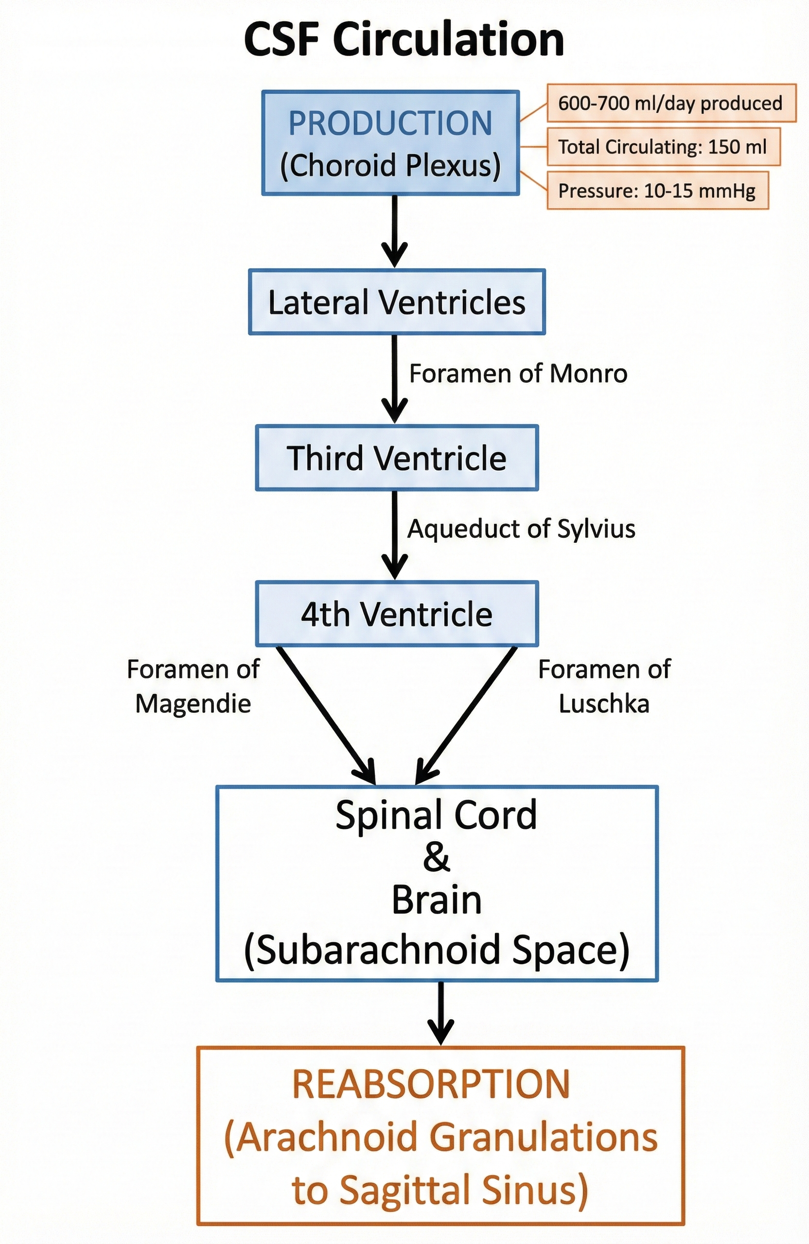
CSF Circulation Pathway — from the choroid plexus through the ventricular system, exiting via the 4th ventricle foramina, circulating through the subarachnoid space, and reabsorbing into the Superior Sagittal Sinus via arachnoid granulations
Mnemonic — How to Remember the Foramina
Both foramina start with their corresponding spatial word:
Foramen of Magendie → Median (single, midline aperture in the roof of the 4th ventricle)
Foramina of Luschka → Lateral (paired, one on each side of the 4th ventricle)
Together they allow CSF to exit into the cisterna magna (Magendie) and the lateral cerebellopontine cisterns (Luschka). Blockage of all three (e.g. in meningitis) causes communicating hydrocephalus.