Skin Anatomy, Physiology & Descriptive Terminology
Structure and function of the skin, its component cells and appendages, and the systematic language used to describe skin lesions
The Epidermis
The epidermis is a stratified squamous epithelium that forms the outermost layer of the skin. It is avascular and relies on diffusion from the underlying dermis for nutrition. Thickness varies considerably: from approximately 0.05 mm on the eyelids to 1.5 mm on the palms and soles.
Layers of the Epidermis — Deep to Superficial
The mnemonic “Come, Let’s Get Sun-Burned” (Corneum, Lucidum, Granulosum, Spinosum, Basale — read superficial to deep) helps recall the five layers. In clinical practice, remember them deep to superficial:
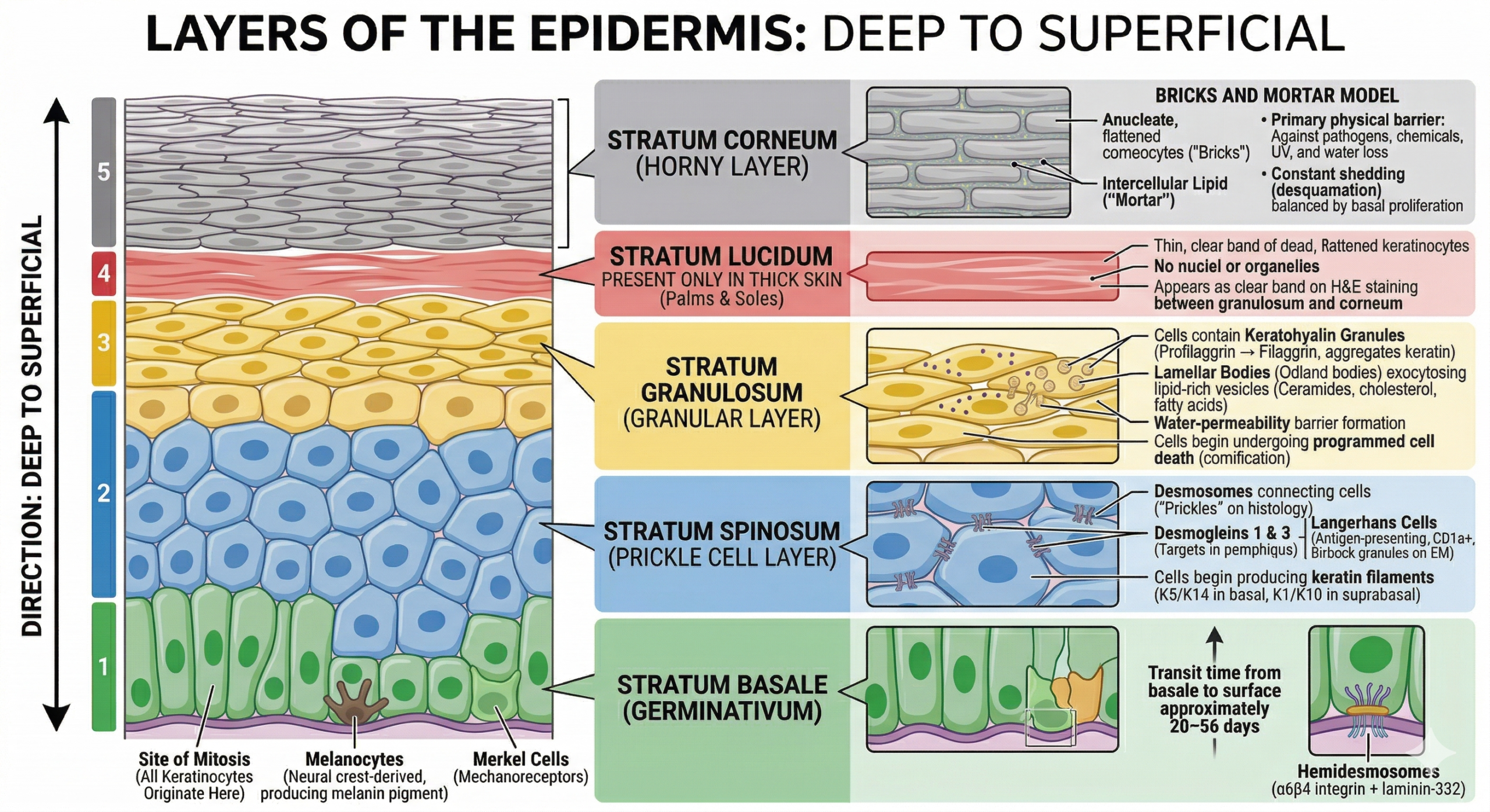
Layers of the epidermis from deep (stratum basale) to superficial (stratum corneum)
Stratum Basale (Germinativum)
Single layer of columnar/cuboidal keratinocytes on the basement membrane. Site of mitosis — all keratinocytes originate here. Contains melanocytes (neural crest–derived) and Merkel cells (mechanoreceptors). Keratinocytes attach to basement membrane via hemidesmosomes (α6β4 integrin + laminin-332). Transit time from basale to surface approximately 28–56 days.
Stratum Spinosum (Prickle Cell Layer)
Several cell layers thick. Cells connected by desmosomes (appear as “prickles” on histology — desmogleins 1 & 3 are key desmoglein targets in pemphigus). Langerhans cells (antigen-presenting, CD1a+, Birbeck granules on EM) reside here. Cells begin producing keratin filaments (K5/K14 in basal, K1/K10 in suprabasal).
Stratum Granulosum (Granular Layer)
2–5 cell layers. Cells contain keratohyalin granules (profilaggrin → filaggrin, which aggregates keratin filaments) and lamellar bodies (Odland bodies) — lipid-rich vesicles that exocytose intercellular lipids (ceramides, cholesterol, fatty acids) forming the skin’s water-permeability barrier. Cells begin undergoing programmed cell death (cornification).
Stratum Lucidum
Present only in thick skin (palms and soles). Thin, translucent, homogeneous layer of dead, flattened keratinocytes. No nuclei or organelles. Appears as a clear band on H&E staining between granulosum and corneum.
Stratum Corneum (Horny Layer)
15–20 layers of anucleate, flattened cornified cells (corneocytes) — “bricks” surrounded by intercellular lipid “mortar.” Corneocytes are filled with keratin filaments bundled by filaggrin. Constant shedding (desquamation) balanced by basal proliferation. Primary physical barrier against pathogens, chemicals, UV, and water loss.
Cornification vs. Keratinisation
Cornification is the terminal differentiation programme of keratinocytes. It involves loss of nucleus and organelles, formation of the cornified envelope (cross-linked structural proteins: involucrin, loricrin, filaggrin), and deposition of intracellular keratin. The result is a metabolically inert, physically resilient barrier cell. Dysregulation leads to conditions such as ichthyosis (failure of desquamation), psoriasis (accelerated turnover — cycle reduced to 4–7 days), and palmoplantar keratodermas.
Epidermal–Dermal Junction (Basement Membrane Zone)
A specialised structure anchoring the epidermis to the dermis, consisting of four zones: plasma membrane of basal keratinocytes → lamina lucida (laminin-332, nidogen) → lamina densa (type IV collagen network) → sub-lamina densa (anchoring fibrils — type VII collagen). Disruption leads to blistering disorders:
| Level of Split | Disorder | Target Antigen |
|---|---|---|
| Intra-epidermal (spinosum) | Pemphigus vulgaris | Desmoglein 3 (±1) |
| Lamina lucida | Bullous pemphigoid | BP180 (collagen XVII), BP230 |
| Sub-lamina densa | Epidermolysis bullosa acquisita | Type VII collagen |