💀 Skull Base & Foramina
A complete, exam-ready guide to every foramen, its contents, its bone, and the clinical consequences of damage. Organised by cranial fossa.
The Skull Base — Your Map of the Territory
The skull base is one of the most heavily tested areas in surgical anatomy. Twelve cranial nerves, the major cerebral arteries, and critical venous channels all thread through specific, named holes — and each one has a clinical story when damaged. The key to mastering this topic is learning the geography first (which fossa, which bone) and then layering on the contents and clinical pearls.
Internal Skull Base — Superior View
The internal surface of the skull base divided into its three cranial fossae: Anterior (frontal), Middle (temporal), and Posterior (occipital). Note how each fossa is at a progressively lower level — like a stepped amphitheatre.
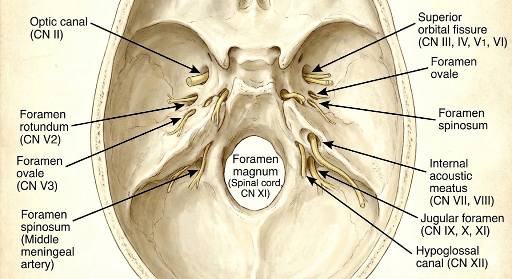
Skull Base Foramina — Annotated Diagram
A labelled overview of the key foramina and fissures of the skull base, colour-coded by cranial fossa. Use this alongside the text to build a spatial mental model of the relationships.
The Three Cranial Fossae — A Framework
Think of the skull base as three stepped terraces descending from front to back, each housing specific parts of the brain and transmitting specific neurovascular structures:
| Fossa | Key Bone(s) | Brain Part Sitting Here | CNs Exiting | Memory Tag |
|---|---|---|---|---|
| Anterior ACF | Frontal, Ethmoid, Sphenoid (lesser wing) | Frontal lobes, Olfactory bulbs | CN I | The “Smell Zone” |
| Middle MCF | Sphenoid (greater wing & body), Temporal (petrous) | Temporal lobes, Pituitary gland | CN II–VI | The “Danger Zone” |
| Posterior PCF | Occipital, Temporal (petrous), Sphenoid (dorsum) | Cerebellum, Brainstem (pons & medulla) | CN VII–XII | The “Downstairs Zone” |
The Master Rule — Which Nerves in Which Fossa?
Anterior Fossa: CN I only
Middle Fossa: CN II, III, IV, V, VI
Posterior Fossa: CN VII, VIII, IX, X, XI, XII
The V (trigeminal) branches each exit through a different foramen in the middle fossa — remember “Standing Room Only”: V1 → Superior Orbital Fissure, V2 → Foramen Rotundum, V3 → Foramen Ovale.
Quick-Reference Master Table
| Foramen / Opening | Bone | Fossa | Key Contents |
|---|---|---|---|
| Cribriform Plate | Ethmoid | ACF | CN I (olfactory filaments) |
| Optic Canal | Sphenoid | MCF | CN II + Ophthalmic Artery |
| Superior Orbital Fissure | Sphenoid | MCF | CN III, IV, V1, VI + Superior Ophthalmic Vein |
| Foramen Rotundum | Sphenoid | MCF | CN V2 (Maxillary) |
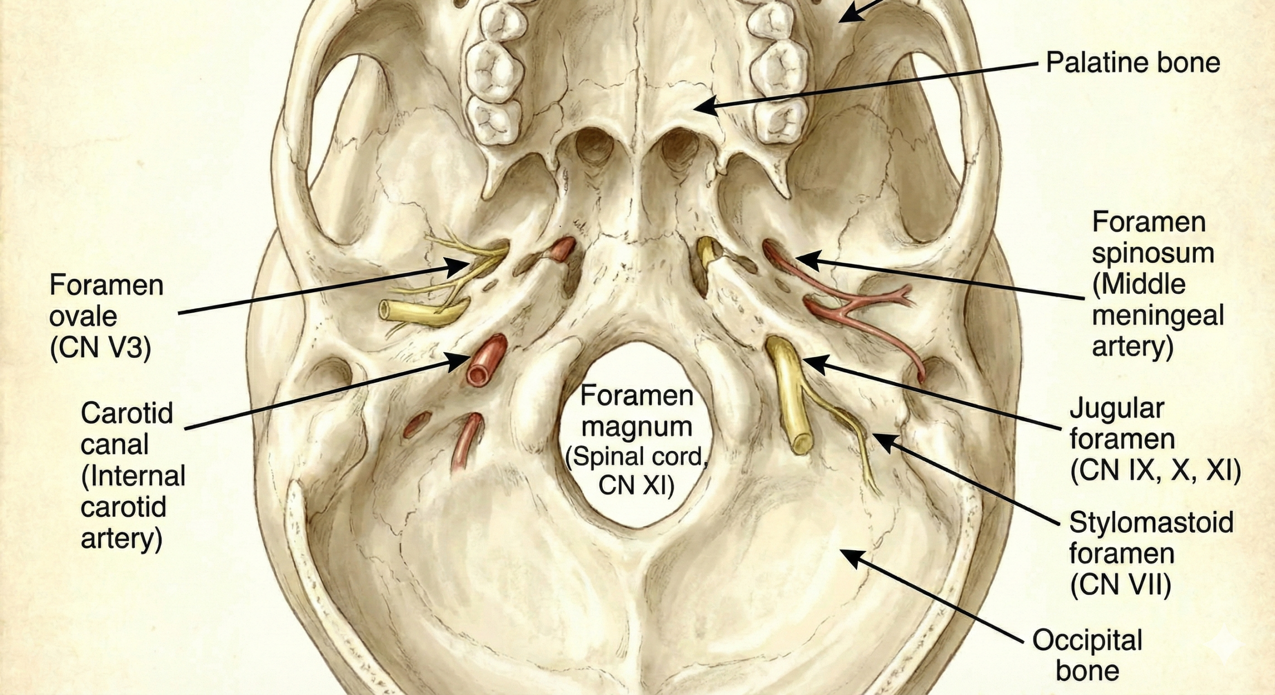
| Foramen Ovale | Sphenoid | MCF | CN V3 + Accessory meningeal a. + Lesser petrosal n. + Emissary v. |
| Foramen Spinosum | Sphenoid | MCF | Middle Meningeal Artery + Nervus Spinosus (V3 meningeal br.) |
| Foramen Lacerum | Sphenoid/Temporal | MCF | ICA passes over it (plugged with cartilage) + Greater Petrosal Nerve |
| Carotid Canal | Petrous Temporal | MCF | Internal Carotid Artery + Sympathetic plexus |
| Internal Acoustic Meatus | Petrous Temporal | PCF | CN VII + CN VIII + Labyrinthine Artery |
| Jugular Foramen | Temporal/Occipital | PCF | CN IX, X, XI + Internal Jugular Vein + Inferior petrosal sinus |
| Hypoglossal Canal | Occipital | PCF | CN XII |
| Foramen Magnum | Occipital | PCF | Medulla → Spinal Cord + Vertebral Arteries + Spinal CN XI roots |